
Quick Take
- Bronchitis inflames the main airways; pneumonia inflames the lung tissue itself.
- Both can be triggered by viruses or bacteria and often follow one another.
- Chest X‑ray and sputum culture are key tools to tell them apart.
- Treatments overlap-antibiotics, bronchodilators, and supportive care-but dosage and duration differ.
- Vaccines, quitting smoking, and early medical attention cut the risk of progression.
Bronchitis is a lower respiratory tract infection that inflames the bronchial tubes, the airways that carry air to the lungs. It can be acute-lasting a few weeks-or chronic, persisting for months or years, especially in smokers or people with COPD.
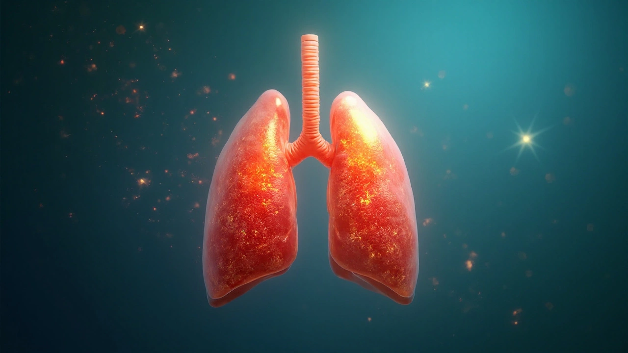
Pneumonia is a lung infection that fills the alveoli (tiny air sacs) with fluid or pus, impairing gas exchange. It may arise on its own or develop after an upper respiratory illness.
What Exactly Is Bronchitis?
Bronchitis belongs to the broader class of lower respiratory tract infections. Its hallmark is a persistent cough that produces sputum-often grey or yellow. The inflamed bronchi produce excess mucus, leading to wheezing, shortness of breath, and chest tightness. Acute bronchitis is usually viral (e.g., rhinovirus, influenza), while chronic bronchitis is linked to long‑term irritants like tobacco smoke or occupational dust.
What Exactly Is Pneumonia?
Pneumonia targets the pulmonary alveoli, the sites where oxygen transfers into the bloodstream. Common culprits include Streptococcus pneumoniae, Haemophilus influenzae, and viral agents such as influenza or RSV. Symptoms overlap with bronchitis-cough, fever, fatigue-but the cough is often more severe, producing thick, sometimes blood‑tinged sputum. Shortness of breath tends to be more pronounced because the alveoli can’t fill with air properly.
How Bronchitis and Pneumonia Are Connected
Think of bronchitis as a road that feeds the lungs. When that road gets clogged with mucus and inflammation, the downstream alveoli become vulnerable. A viral or bacterial agent that first irritates the bronchi can quickly spread deeper, turning an episode of bronchitis into pneumonia. This progression is especially common in:
- Older adults with weakened immune systems.
- People with chronic lung diseases (COPD, asthma).
- Smokers whose cilia-tiny hair‑like cleaners-are damaged.
Studies from the American Thoracic Society show that up to 30% of hospitalized acute bronchitis cases develop secondary bacterial pneumonia within two weeks if left untreated.
Shared Risk Factors and Warning Signs
Both conditions share a set of risk factors that clinicians use to gauge the likelihood of escalation:
| Factor | Impact |
|---|---|
| Smoking | Damages airway cilia, reduces clearance |
| Advanced age (≥65) | Immune response slows |
| Chronic lung disease | Baseline inflammation primed for infection |
| Recent viral upper‑respiratory infection | Creates a breeding ground for bacteria |
| Immunosuppression (meds, HIV) | Limits ability to fight pathogens |
Key warning signs that bronchitis may be morphing into pneumonia include high fever (>38.5°C), rapid breathing, chest pain that worsens with deep breaths, and a sudden drop in oxygen saturation (below 92%). If any of these appear, a doctor should order imaging.

Diagnosing the Overlap: Imaging and Lab Tests
Chest X‑ray remains the gold‑standard for distinguishing bronchial inflammation from alveolar infiltrates. In bronchitis, the X‑ray is usually clear; in pneumonia, you’ll see patchy opacities or consolidation.
Additional tools:
- Sputum culture: Identifies bacterial pathogens, guiding antibiotic choice.
- Blood tests: Elevated white‑blood‑cell count hints at bacterial involvement.
- Pulse oximetry: Detects hypoxia early, especially in elderly patients.
When Both Conditions Co‑Exist: Treatment Strategies
Therapy aims to clear the infection, reduce inflammation, and support breathing.
- Antibiotics: Reserved for confirmed bacterial cases or high‑risk patients. Common regimens include amoxicillin‑clavulanate for Streptococcus pneumoniae or a macrolide for atypical organisms.
- Bronchodilators: Short‑acting beta‑agonists (e.g., salbutamol) open the bronchial tubes, easing cough and wheeze.
- Corticosteroids: Low‑dose oral steroids can reduce airway swelling in severe bronchitis, but are used cautiously in pneumonia due to infection‑risk concerns.
- Supportive care: Hydration, rest, and oxygen therapy for hypoxic patients.
Duration matters: acute bronchitis often resolves in 7‑10days, while bacterial pneumonia typically requires 5‑7days of antibiotics after the fever subsides.
Prevention: Cutting the Path from Bronchitis to Pneumonia
Vaccines are the single most effective barrier. The annual influenza vaccine reduces viral bronchitis cases, while the pneumococcal vaccine guards against the most common bacterial pneumonia strain.
Lifestyle tweaks further lower risk:
- Quit smoking - mucociliary clearance rebounds within weeks.
- Maintain good hand hygiene during flu season.
- Manage chronic conditions (diabetes, heart disease) with regular check‑ups.
- Stay active - aerobic exercise improves lung capacity.
Related Concepts and Next Steps
Understanding the bronchitis‑pneumonia link opens doors to deeper topics such as:
- Chronic Obstructive Pulmonary Disease (COPD) - how long‑term airway damage predisposes to infections.
- Asthma exacerbations - overlapping symptoms and treatment nuances.
- Community‑acquired vs. Hospital‑acquired pneumonia - differences in pathogen profile and antibiotic stewardship.
- Respiratory physiotherapy - techniques to clear mucus and improve ventilation.
Readers who mastered the basics may want to explore diagnostic algorithms (e.g., CURB‑65 scoring) or delve into the latest antimicrobial guidelines from the Infectious Diseases Society of America.
Quick Comparison: Bronchitis vs Pneumonia
| Aspect | Bronchitis | Pneumonia |
|---|---|---|
| Primary Site | Bronchial tubes (airways) | Alveoli (lung tissue) |
| Typical Cause | Viruses (70%); bacteria (30%) | Bacteria (most common), viruses, fungi |
| Main Symptom | Productive cough, mild wheeze | Fever, chills, chest pain, productive cough |
| Chest X‑ray | Usually clear | Shows infiltrates or consolidation |
| Treatment Focus | Bronchodilators, fluids, sometimes antibiotics | Targeted antibiotics, oxygen, possible steroids |
Frequently Asked Questions
Can bronchitis turn into pneumonia?
Yes. When the infection spreads from the bronchi to the alveoli, the inflammation deepens and pneumonia develops. This is more common in older adults, smokers, and people with chronic lung disease.
How do doctors differentiate between the two?
A clear chest X‑ray points to bronchitis, while visible infiltrates indicate pneumonia. Sputum analysis and blood tests also help, especially if bacterial infection is suspected.
Is antibiotics always needed?
Not for viral bronchitis or viral pneumonia. Antibiotics are reserved for confirmed bacterial cases or high‑risk patients where the benefit outweighs resistance concerns.
What vaccines protect against these illnesses?
The annual flu shot reduces viral bronchitis, and the pneumococcal vaccine (PCV13 or PPSV23) guards against the most common bacterial cause of pneumonia.
When should I seek emergency care?
If you experience rapid breathing, persistent high fever, chest pain that worsens with inhalation, or oxygen saturation below 92%, go to the ER immediately. These signs often signal pneumonia or severe bronchial obstruction.
Our healthcare system must prioritize preventing these infections to keep the nation strong.
Great rundown on how bronchitis can set the stage for pneumonia 😊
Keeping an eye on those warning signs is crucial for anyone dealing with a cough that just won’t quit.
Vaccines are a solid frontline, especially the flu shot and pneumococcal vaccine.
Quitting smoking not only improves cilia function but also reduces the chance of a secondary infection.
Stay hydrated and rest, because your body needs every ounce of energy to fight off the microbes.
The distinction between clear X‑rays in bronchitis and infiltrates in pneumonia is essential.
Early sputum cultures guide appropriate antibiotic use.
People keep overusing antibiotics for simple viral bronchitis.
While the layperson may skim the headings, a deeper appreciation of the pathophysiological cascade demands a more rigorous deconstruction; indeed, the progression from bronchial mucosal inflammation to alveolar exudation is not merely a linear escalation, but a complex interplay of host defenses and microbial virulence factors.
First, consider the epithelial barrier: in acute bronchitis, the ciliated epithelium remains largely intact, albeit besieged by excess mucus, whereas in pneumonia the alveolar-capillary membrane suffers from endothelial leakage and surfactant disruption.
Second, the immune response diverges; neutrophil migration predominates in bacterial pneumonia, producing the characteristic purulent sputum, while viral bronchitis may elicit a lymphocytic infiltrate that resolves spontaneously.
Third, diagnostic imaging underscores this dichotomy: a clear radiograph signifies isolated bronchial involvement, whereas patchy infiltrates or consolidation unequivocally point to alveolar participation.
Moreover, the therapeutic regimen must be calibrated accordingly: bronchodilators and supportive fluids suffice for most bronchitic episodes, yet pneumonia obliges targeted antimicrobial therapy, often guided by sputum culture and sensitivity patterns.
It is also incumbent upon clinicians to judiciously assess severity scores such as CURB‑65, which stratify mortality risk and inform inpatient versus outpatient management.
Furthermore, the role of adjunctive corticosteroids remains contentious; low‑dose steroids can mitigate airway edema in severe bronchitis, but their immunosuppressive potential may exacerbate bacterial proliferation in pneumonia.
Preventive strategies, including the annual influenza vaccine and pneumococcal immunization, serve as the cornerstone of public health interventions, dramatically curtailing incidence rates across demographic strata.
Smoking cessation, beyond restoring mucociliary clearance, also reduces oxidative stress that predisposes the pulmonary parenchyma to infection.
In the elderly, vigilant monitoring of oxygen saturation is paramount, as hypoxemia may herald an occult transition from bronchitis to pneumonia.
Clinical vigilance, therefore, hinges on recognizing subtle shifts: escalating fever, tachypnea, pleuritic chest pain, and a precipitous drop in SpO₂.
In summary, appreciating the nuanced continuum between bronchial and alveolar pathology empowers clinicians to tailor diagnostics, optimize antimicrobial stewardship, and ultimately improve patient outcomes.
From a motivational standpoint, think of your respiratory system as a high‑performance engine; regular maintenance-like vaccinations and smoking cessation-keeps the airflow smooth and prevents catastrophic shutdowns.
The jargon here-ciliary clearance, alveolar infiltrates, CURB‑65-might sound intimidating, but they’re simply tools that help us gauge risk and act decisively.
If you notice the cough lingering beyond a week with colored sputum, that’s a signal to consider a deeper evaluation.
Remember, early intervention isn’t just about feeling better faster; it’s about averting the cascade that turns a manageable bronchial irritation into a life‑threatening pneumonia.
Contemplating the thin veil between irritation and infection invites a philosophical musing: where does symptom end and disease begin?;
Perhaps the answer lies not in the lungs alone, but in the collective vigilance of those who heed the subtle cues of their own bodies.